
Fundus Photography
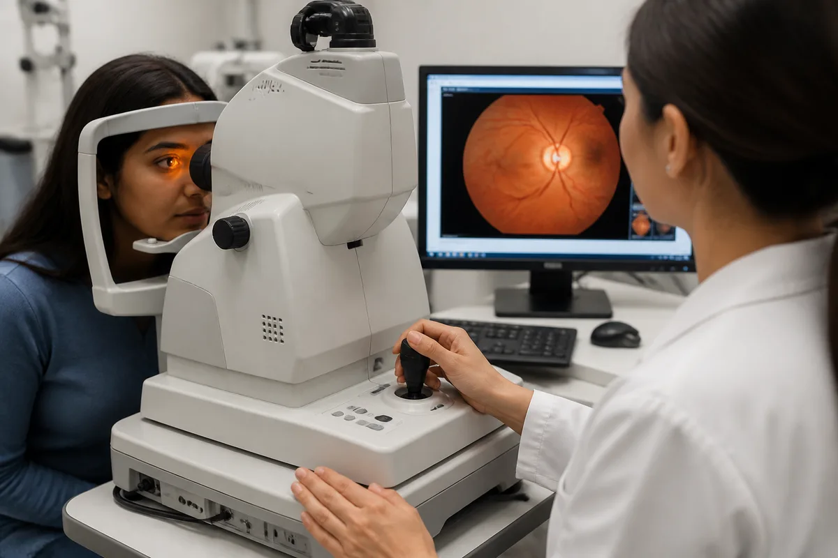
Fundus Photography
Fundus photography documents the retina and optic nerve in high resolution, creating a record that can be compared precisely at future visits.
What we document
- Optic nerve appearance — the baseline that glaucoma monitoring depends on.
- Retinal changes from diabetes and blood pressure — findings reported back to your medical providers.
- Moles and lesions of the retina — watched over time for any change.
Patients see their own images during the exam, which makes the explanation of any finding considerably clearer.
